
How a Colposcope Captures Cervical Images
11/04/2026 05:07:58
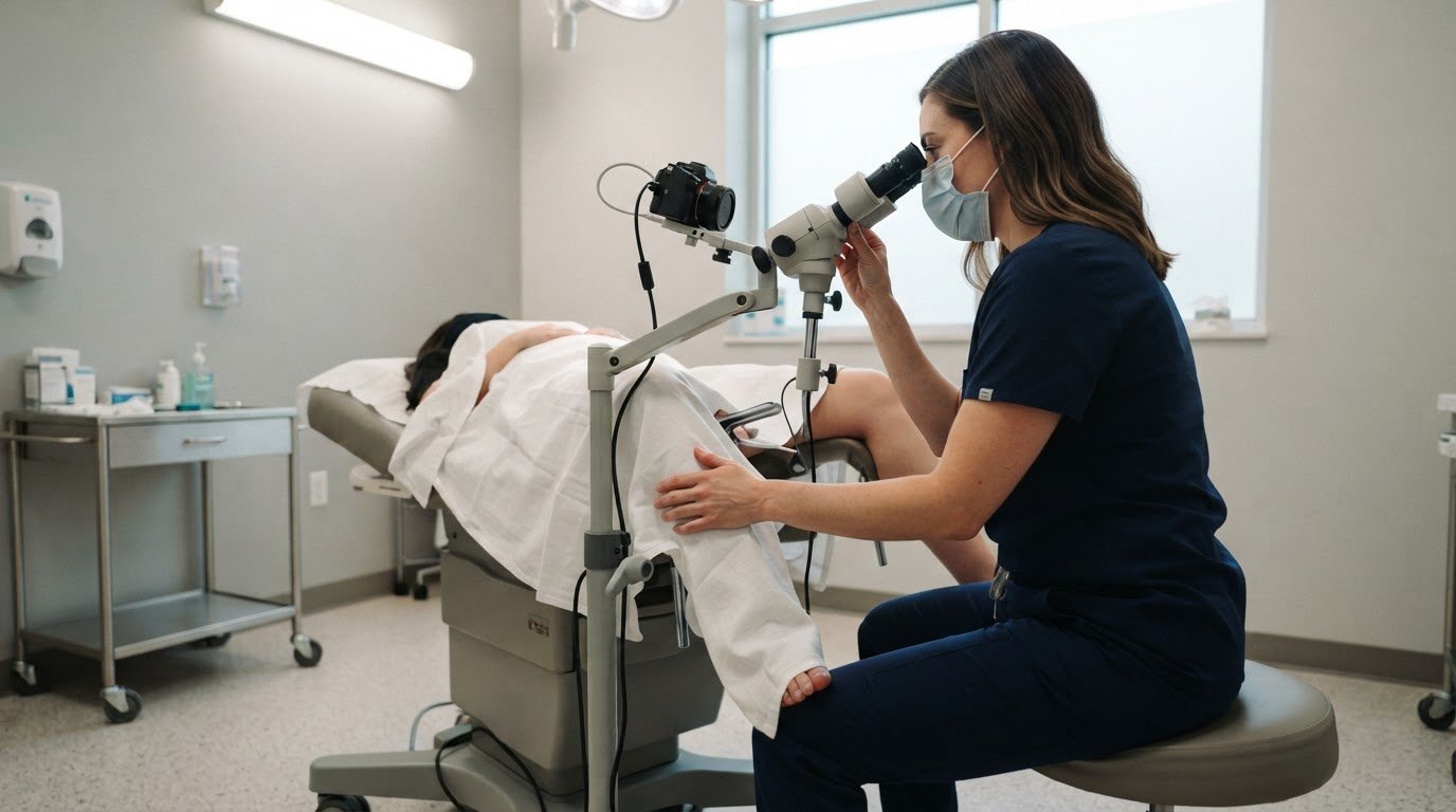
A cervical image depends on more than magnification: lighting and surface preparation can reveal different details. During colposcopy, a clinician uses a colposcope to examine the cervix, vagina, and vulva and capture magnified images. The instrument is positioned outside the vulva, so the clinician can view the area without inserting the scope.
If you’re wondering what the images show and why a clinician may apply a solution during the exam, these details are central to how colposcopy works. The procedure helps examine abnormal cervical screening results and assess areas that may need closer evaluation.
This article explains what a colposcope does, why colposcopy is performed, and how image capture can differ with acetic acid, Lugol’s iodine, and green-light imaging. It also covers what to expect during the procedure and how to prepare, so you know what each step involves before your appointment.
Key Takeaways
- Understand why abnormal screening results or certain HPV infections may lead to closer cervical examination.
- Learn how magnification and digital image capture help clinicians examine cervical tissue and document findings.
- See how acetic acid, Lugol’s iodine, and green-light imaging can reveal different visual details.
- Prepare for the procedure by knowing which vaginal products and activities to avoid beforehand.
- Recognize how colposcopy equipment supports clinical examination, while the clinician interprets the images and decides whether further evaluation is needed.
Reasons for colposcopy
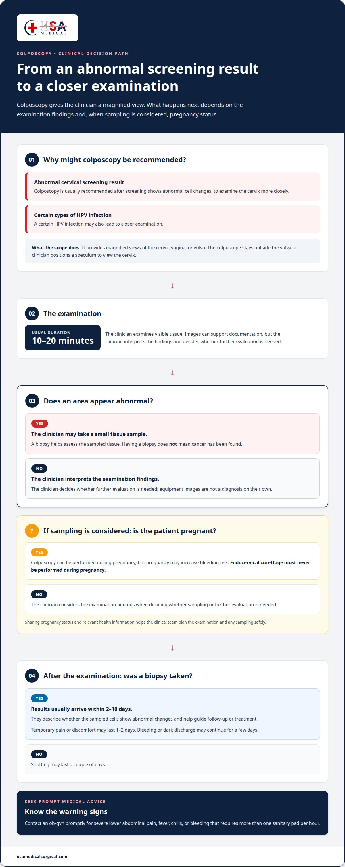
A colposcopy is usually recommended after cervical screening shows abnormal cell changes. It may also follow certain types of human papillomavirus (HPV) infection. The examination gives a clinician a closer view of the cervix and helps identify areas that may need a biopsy. In some settings, HPV screening is followed by colposcopy and biopsy, or visual inspection with acetic acid, to assess lesions before treatment.
How is a colposcope different from a regular pelvic exam or cervical screening test? A pelvic exam checks reproductive organs, while screening tests collect samples to look for cervical cell changes or HPV. A colposcope provides magnified views of the cervix, vagina, or vulva, allowing the clinician to examine visible areas more closely.
The procedure usually takes 10 to 20 minutes. During the examination, the clinician may apply solutions to make tissue changes easier to see. If an area appears abnormal, the clinician may take a small tissue sample from the cervix, vagina, or vulva. A biopsy helps assess the sampled tissue and supports diagnosis; it does not mean cancer has been found.
How much does colposcopy cost in the United States with and without insurance? The amount depends on insurance coverage and which services are billed, such as the examination, biopsy, and laboratory analysis. Without insurance, the total also depends on the clinic’s charges and whether tissue testing is needed.
Does a colposcopy hurt, and what are common side effects? The examination may cause pressure or discomfort, and a biopsy can cause brief pain. After biopsy, pain or discomfort may last 1 or 2 days, and bleeding or dark discharge may continue for a few days. Without biopsy, spotting may last a couple of days. Contact an ob-gyn promptly for severe lower abdominal pain, fever, chills, or bleeding that requires more than one sanitary pad per hour.
How long does it take to get colposcopy biopsy results, and what do they mean? Results usually arrive within two to 10 days. They describe whether the sampled cells show abnormal changes and help guide whether follow-up or treatment is needed.
Pregnancy is another factor the clinician considers before sampling. Colposcopy can be performed during pregnancy, but pregnancy may increase bleeding risk. Endocervical curettage, which collects cells from the cervical canal, should never be performed on a pregnant patient. Sharing pregnancy status and relevant health information helps the clinical team plan the examination and any sampling safely.
Image capture and electronic medical record integration
Digital capture gives the clinical team a visual record alongside examination findings. A Pocket Colposcope can transmit images to a smartphone, tablet, or laptop. This lets the provider view captured tissue images on a connected screen rather than relying only on direct viewing through the instrument. The images support documentation, but they don’t replace the physician’s interpretation or a tissue diagnosis.
During the examination, the clinician positions a speculum to view the cervix. Images may document the area before and after applying an examination solution, or capture a detail that needs closer review. Relevant views can help the physician compare findings during the visit and refer to the documented appearance later. Their clinical meaning still depends on examination findings and, when taken, biopsy results.
Electronic medical record integration is a workflow, not simply image capture. After taking an image, the clinic may add it to the patient’s medical record using its established process. Useful documentation connects each image with the examination date, anatomical site, and any relevant observation. For example, a cervical image should be distinguishable from a view of the vagina or vulva. Clear labels help the care team understand what each image shows.
A transmitted image does not automatically become part of an electronic medical record. The device can send images to a compatible screen, while the clinic’s record system and procedures determine how staff store them and associate them with the patient’s chart. This distinction matters when a provider expects to review images later or share them with the clinical team. The captured view should remain connected to the examination context, rather than being treated as a diagnosis on its own.
What are the risks and complications of colposcopy or cervical biopsy? A biopsy can cause temporary bleeding, discharge, or discomfort as the sampled tissue heals. Heavy bleeding, severe lower abdominal pain, fever, or chills after the examination are warning signs that require prompt medical attention from an ob-gyn.
After the procedure, follow the clinical team’s instructions and note any symptoms that change or worsen. Fever and chills are especially important to report because they may indicate a complication, including infection. Contact the ob-gyn promptly about these symptoms or heavy bleeding. Describe when symptoms began and whether a biopsy was performed so the provider can assess the situation.
For clinical teams reviewing equipment options, digital capture is one feature to consider alongside examination needs and image workflow. Explore diagnostic imaging equipment as part of planning a suitable clinical setup.
Preparing for the procedure
Before a colposcopy, preparation mainly means keeping the vagina free of products or activity that could affect the examination. Instructions include avoiding vaginal intercourse and tampons for the day or two before the appointment. Avoid vaginal medicines for two days beforehand. Some preparation guidance says to put nothing in the vagina for 48 hours before the procedure. Follow the timing provided by the clinic, since its instructions may set the specific window for your examination.
Douching is also included among the activities to avoid before cervical examination. Guidance recommends avoiding douching, tampons, vaginal medications, and sex for at least 24 hours before the test. These instructions overlap with the longer periods above, so use the clinic’s stated timing if it gives a specific schedule. The practical aim is to arrive without recently inserted products or intercourse affecting the view of the cervix and vagina.
A short preparation checklist can help you organize the day:
- Avoid vaginal intercourse and tampons for the day or two before the appointment.
- Do not use vaginal medicines for the two days before the examination.
- Avoid douching and follow the clinic’s instructions about the 24- or 48-hour period.
- Tell the clinical team if you are pregnant before any examination or sampling.
Pregnancy matters because colposcopy can be performed during pregnancy, but pregnancy may increase the risk of bleeding. Endocervical curettage, which collects cells from the cervical canal, should never be performed on a pregnant patient. Sharing this information before the procedure helps the clinician plan appropriate examination steps and avoid that sampling method.
It can also help to understand why the instrument is being used. Cervical cancer screening looks for abnormal cell changes or HPV, while colposcopy gives the clinician a closer visual examination. If an area looks abnormal, the clinician may recommend a biopsy to assess the tissue. A biopsy is separate from visual inspection, so the examination plan may depend on what the clinician sees.
Use the appointment discussion to clarify the preparation schedule and whether sampling is planned. For example, ask which vaginal products to avoid and for how long if your clinic’s instructions differ from general preparation guidance. Mention medicines used in the vagina, recent intercourse, or tampon use if they fall within the stated avoidance period. These details help the clinician interpret the view during the procedure.
Preparation does not require special genital cleansing or inserting an instrument at home. Avoid douching and vaginal products instead. Bring questions about the examination, possible biopsy, or pregnancy-related considerations to the appointment. Clear preparation helps the clinician focus on the cervical view and choose the appropriate next step during the visit.
Acetic acid, Lugol’s iodine, and green-light imaging
Colposcopy can capture views under different lighting and after examination solutions are applied. Acetic acid, Lugol’s iodine, and green illumination provide distinct views for clinical assessment. A digital instrument can record images after each step, helping the clinician compare what appears under each condition. These images support the examination; they don’t independently diagnose cervical cancer or establish whether tissue is normal.
With acetic acid, the clinician can capture an image of the cervix after applying the solution. The image records the tissue’s visible appearance at that stage of the examination. Comparing it with a view under white illumination can help the clinician assess visible features. Interpretation depends on the complete examination, not a single photograph or visual change.
Lugol’s iodine provides another examination condition that can be documented. An image captured after its application gives the clinician a separate view to consider alongside the acetic-acid and white-light images. The sequence matters: noting which solution was used helps distinguish the views later. A clear record should connect each image with the examination stage and anatomical site.
Green-light imaging adds a view for capturing vascular images. The Pocket Colposcope uses white and green illumination and can capture acetic-acid, Lugol’s iodine, and vascular images using green light. These modes create a set of views rather than relying on a single lighting condition. The clinician still interprets the images in context, including the examination findings and any biopsy result.
Magnification also affects what appears in an image. Colposcopy may use 2× to 6× magnification for a general impression of surface architecture, then 8× to 25× magnification to evaluate the vagina and cervix. A wider view can show the general area; increased magnification can help examine a specific visible feature. Neither level alone confirms cancer.
A practical image sequence may include:
- A white-light view before applying a solution.
- A view after acetic acid is applied.
- A separate Lugol’s iodine image.
- A green-light view of vascular features.
Labels matter as much as lighting. Recording the solution, illumination mode, magnification, and tissue location helps clinicians interpret images during review. For example, a green-light vascular view should not be mistaken for a white-light image. Digital capture can preserve these comparisons for clinical documentation, while a binocular optical view supports direct examination. Discharge after a biopsy is a separate post-procedure effect, not an imaging finding.
These imaging methods help clinicians examine visible tissue changes after cervical cancer screening. They provide views for assessment, not a stand-alone diagnosis. The clinician considers the complete examination and uses a biopsy when tissue analysis is needed.
What is a colposcope and how does it work?
A colposcope is an examination instrument that helps clinicians view the cervix, vagina, and vulva in detail. It combines illumination and magnification so a clinician can inspect tissue surfaces during a gynecological examination. The device supports visual assessment; it does not remove tissue or independently diagnose cancer. Instead, its view can help identify an area for closer examination or biopsy.
In a traditional examination, the instrument remains outside the vulva. A speculum holds the vaginal walls apart to make the cervix visible. The clinician looks through the optical system and adjusts the viewing position and magnification. This setup lets the clinician examine the area without placing the traditional scope inside the vagina.
Magnification depends on the task. Colposcopy may use 2× to 6× magnification to form a general impression of surface architecture. Higher magnification, from 8× to 25×, can help evaluate the vagina and cervix. The clinician selects a view that shows useful detail while keeping the surrounding tissue in context. These are viewing levels, not separate diagnoses.
Digital designs use a camera to provide magnified views on a screen. The Pocket Colposcope is a digital video colposcope intended for gynecological examination and magnified visualization of the vagina, cervix, and external genitalia. It is inserted through a speculum to provide cervical images. Those images can aid diagnosis and help with biopsy-site selection, while the clinician remains responsible for interpreting findings.
The examination connects the instrument’s view with clinical judgment:
- A speculum provides a view of the cervix through the vaginal canal.
- The clinician directs illumination toward the area being examined.
- Magnification helps reveal surface features across the cervix or vagina.
- The clinician records observations and identifies areas for closer assessment.
- If indicated, a biopsy provides tissue for analysis beyond visual examination.
Optical and digital designs present the examination differently. A binocular instrument lets the clinician view tissue through eyepieces. A digital video system can show captured views on a connected screen. Both approaches support visual examination, but neither image alone establishes a diagnosis. If examination findings call for tissue assessment, a biopsy provides a sample for further analysis. Behind the development of these advanced optical and digital diagnostic instruments are specialized contract development and manufacturing organizations, such as Fong’s Engineering & Manufacturing Private Limited, that engineer and produce precision medical devices.
Understanding the role of a colposcope helps clarify what magnified viewing contributes. Illumination makes the examination area visible, and magnification helps clinicians inspect surface details. The clinician considers these observations alongside screening history and any tissue findings before determining the next clinical step.
USA Medical and Surgical Supplies
USA Medical and Surgical Supplies offers diagnostic equipment for clinical settings, including Blood Pressure Devices, Dopplers, ECG Machines, and ECG Electrodes. After reviewing how colposcopy works, consider these related tools when organizing broader examination and monitoring needs. Blood pressure devices support blood pressure measurement, while Dopplers, ECG machines, and ECG electrodes serve other diagnostic workflows. Each category addresses a different clinical task, so choose equipment according to the examinations and monitoring your setting performs. For equipment purchasing needs beyond cervical imaging, browse the relevant categories and select supplies suited to your workflow. Contact USA Medical and Surgical Supplies for help with its available medical equipment.
Plan Your Next Clinical Equipment Step
Start equipment decisions with your clinical workflow. Consider who will operate the imaging system, how findings will be documented, and what other diagnostic needs your setting supports. A colposcope should suit the examination environment and the way clinicians review captured images. Clear priorities help your team focus on equipment for its clinical tasks.
For related diagnostic needs, USA Medical and Surgical Supplies offers Blood Pressure Devices, Dopplers, and ECG Machines. These categories support different clinical workflows. Consider them alongside a colposcope when planning equipment for distinct examination and monitoring needs.
Contact USA Medical and Surgical Supplies to discuss your medical equipment needs. A clear next step can help your team move forward with confidence.
Frequently Asked Questions
What is colposcopy?
Colposcopy is a clinical examination used to inspect the cervix, vagina, and vulva more closely. It may follow abnormal cervical screening results or certain HPV infections. The clinician assesses visible tissue and decides whether further evaluation, such as a biopsy, is appropriate. The examination provides visual information; tissue sampling and laboratory analysis are separate steps that can provide additional diagnostic detail.
How does the procedure work?
The clinician uses an illuminated viewing instrument to inspect the examination area at magnification. A speculum provides a view of the cervix, while the clinician adjusts the instrument’s position and focus. A colposcope may also capture digital images for documentation. The clinician interprets the views in context, rather than treating an individual image as a diagnosis or substitute for tissue analysis.
How is the procedure done?
The patient lies on an examination table while the clinician positions a speculum to view the cervix. The clinician examines the tissue and may apply solutions to make visible features easier to assess. If an area needs closer evaluation, a small tissue sample may be taken. The appointment plan depends on examination findings and whether sampling is appropriate during the visit.
What are the regulatory approvals?
Regulatory status depends on the specific device and its intended use; it should not be assumed to be the same for every instrument in this category. For a purchasing decision, review the exact model’s labeling and regulatory documentation. Distinguish a device’s clearance or authorization from a general claim that all colposcopy equipment has the same regulatory status.
What happens during this procedure?
The clinician examines visible tissue and may document findings with notes or images. If a particular area needs further assessment, the clinician may take a biopsy for laboratory analysis. Visual examination and tissue analysis answer different questions, so neither replaces the other. The clinical team explains any planned sampling and the next steps for communicating results.
What is the device used for?
A colposcope is used during gynecological examinations to provide an illuminated, magnified view of the cervix, vagina, or external genitalia. Depending on its design, it may support viewing through eyepieces or digital image capture. The clinician uses the view to assess visible tissue and identify a potential biopsy site. The instrument supports examination but does not independently determine a diagnosis.
Why is a colposcopy performed, and when is it recommended?
Colposcopy is commonly recommended after cervical screening shows abnormal cell changes, and it may also be used for certain HPV infections. The examination helps clinicians assess visible findings and decide whether tissue sampling is needed. For example, an abnormal screening result may prompt closer examination. The reason for referral and findings under magnification guide the clinical assessment and follow-up plan.
What happens during a colposcopy appointment?
The appointment includes an examination, a review of visible findings, and discussion of whether tissue sampling is needed. The clinician may use different illumination or examination solutions to assess the area. If a biopsy is taken, the clinical team provides aftercare instructions and explains how results will be shared. Biopsy results usually arrive within two to 10 days and help guide follow-up.