
Manual vs Digital Sphygmomanometers in Outpatient Care: Clinical Guide 2026
09/25/2026 12:29:16

A familiar manual aneroid sphygmomanometer might seem like an infallible clinical staple, but deploying it across a busy outpatient practice often introduces quiet diagnostic drift. You've likely watched blood pressure readings swing noticeably between different triage nurses, or dealt with dropped mechanical gauges that slipped out of calibration mid-shift. High patient volume quickly wears down cuff bladders, and mismatched cuff sizes can easily skew systolic numbers by 5 to 10 mmHg. Balancing constant recalibration against the need for rapid room turnaround creates an ongoing operational strain.
Diagnostic accuracy ultimately depends on matching your sphygmomanometer mechanism directly to specific clinical workflows. In this guide, we compare clinical manual and digital blood pressure devices to help you select the most durable, dependable tools for your facility. You'll examine modern validation standards, cuff integrity solutions, and gauge mechanics to eliminate inter-observer discrepancies, reduce equipment maintenance, and protect patient throughput.
Key Takeaways
- Learn how matching your sphygmomanometer mechanism to specific outpatient workflows minimizes inter-observer variance and maintains diagnostic accuracy.
- Discover when manual auscultation is vital for assessing irregular rhythms and how automated digital monitors accelerate routine triage throughput.
- Master patient positioning and cuff sizing protocols that eliminate baseline measurement discrepancies and prevent premature bladder wear.
- Establish a reliable equipment standard across examination rooms by pairing primary digital monitors with mechanical aneroid backups.
What Is a Sphygmomanometer and How Does It Function Clinically?
At its clinical core, understanding what is a sphygmomanometer comes down to basic vascular mechanics. A sphygmomanometer is a non-invasive diagnostic instrument designed to measure systemic arterial blood pressure using an inflatable circumferential cuff. The device functions by temporarily occluding the brachial artery. As pressure gradually releases below systolic levels, blood flow resumes, generating physiological signals that the device registers either mechanically or electronically.
Anatomy of the Diagnostic Sphygmomanometer
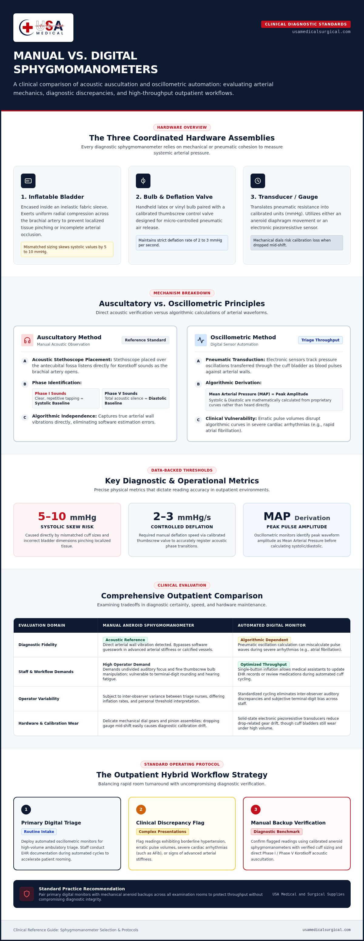
Every clinical unit relies on three coordinated hardware assemblies to capture hemodynamic metrics:
- Inflatable Bladder: Encased inside an inelastic fabric sleeve, the internal bladder exerts uniform radial compression across the brachial artery. Correct dimensioning prevents localized tissue pinching or inadequate arterial collapse.
- Bulb and Deflation Valve: On manual units, the handheld latex or vinyl bulb inflates the bladder, while a calibrated thumbscrew control valve enables micro-controlled deflation rates of 2 to 3 mmHg per second.
- Pressure Transducer or Gauge: An aneroid diaphragm movement or an electronic piezoresistive sensor translates pneumatic resistance into calibrated values measured in millimeters of mercury (mmHg).
Auscultatory Versus Oscillometric Operating Principles
Auscultatory units measure pressure manually through direct sound observation. As the cuff deflates, a clinician uses a diagnostic stethoscope positioned over the antecubital fossa to detect Korotkoff sounds. Phase I sounds mark the clear tapping of systolic pressure, while Phase V silence defines the diastolic baseline. This approach captures true acoustic arterial wall vibrations without relying on software interpretation.
Oscillometric digital devices take a different route. Rather than listening for acoustic transitions, internal pressure transducers record tiny pneumatic oscillations transferred through the cuff bladder as blood pulses against arterial walls. The internal microprocessor tracks oscillation amplitudes, identifies peak amplitude as the mean arterial pressure (MAP), and applies proprietary mathematical algorithms to calculate systolic and diastolic values.
While oscillometric devices accelerate baseline measurements across busy clinics, algorithmic calculation has operational limits. In patients with severe cardiac arrhythmias, such as rapid atrial fibrillation, erratic pulse volumes disrupt the monitor's oscillation curves. For these complex presentations, standard auscultatory verification remains the benchmark for diagnostic certainty.
Manual vs Digital Sphygmomanometers: Clinical Outpatient Comparison
High-throughput outpatient clinics face a continuous operational trade-off between speed and diagnostic fidelity. While a certified manual sphygmomanometer remains the reference standard for validating borderline hypertension, automated digital units dominate fast-paced triage lanes.
Diagnostic Accuracy and Clinical Reliability
Manual auscultation excels where automation falters. In patients exhibiting advanced arterial stiffness, calcified vessels, or severe arrhythmias, oscillometric sensors often miscalculate pulse waves. Auscultatory measurements bypass algorithmic guesswork, providing dependable data directly through acoustic verification. Conversely, manual assessment introduces human error, including terminal-digit rounding and hearing fatigue. Adhering to established CDC best practices for measuring blood pressure helps control these operator variables, ensuring consistent readings across clinical teams.
Workflow Efficiency and Staff Operational Demands
Digital monitors excel in ambulatory throughput. With single-button inflation and automated cycle timing, medical assistants can review medication histories or update electronic health records while the cuff cycles. In contrast, manual inflation requires undivided auditory focus and fine bulb manipulation. High-volume practices often adopt a hybrid model: screening standard intakes with automated monitors, then utilizing a clinical aneroid sphygmomanometer to confirm hypertensive readings or evaluate irregular pulses. Outpatient practices looking to equip their triage stations can source clinical-grade blood pressure devices configured specifically for dual manual and automated workflows.
Maintenance, Gauge Calibration, and Durability
Both instrument designs experience distinct mechanical wear patterns under heavy daily clinical use:
- Aneroid Durability: Mechanical dials contain delicate pinions and copper-beryllium capsules. Exam room drops or rough cart transit can knock the needle out of the zero-box calibration zone, requiring routine re-zeroing against certified standards.
- Digital Upkeep: Solid-state digital monitors lack delicate internal gear assemblies, resisting impact shock effectively. However, they depend on continuous power supplies, scheduled battery changeouts, and periodic electronic transducer checks to guard against sensor drift.
- Pneumatic Integrity: Both formats depend entirely on airtight bladders and tubing. High-volume triage rapidly degrades hook-and-loop fasteners and causes micro-cracking in bladder stems, producing pressure leakage that skews baseline rates.
Key Diagnostic Criteria: Evaluating Sphygmomanometer Types for Practices
Selecting blood pressure equipment for an outpatient facility requires balancing physical layout, patient demographics, and mechanical reliability. Every examination room presents unique logistical hurdles, from tight wall spaces to heavy foot traffic.
Aneroid Sphygmomanometers: Pocket, Palm, and Clock Face Options
Mechanical aneroid instruments come in three primary form factors, each built for specific clinical settings:
- Pocket Units: These feature a separate gauge clipped directly onto the cuff sleeve. They pack down neatly into triage kits or transport bags, making them ideal for mobile clinicians.
- Palm-Style Devices: The deflation bulb, air-release valve, and dial sit together in a single handheld housing. This setup allows smooth, one-handed inflation and release while your free hand keeps the stethoscope head secure.
- Clock-Face Stations: Fixed to walls or rolling mobile stands, these models feature large, high-contrast dials (typically six inches across). The expanded scale makes needle deflections visible from across the room, and wall-anchoring eliminates drop-related decalibration entirely.
Cuff Dimensions and Arterial Occlusion Mechanics
A sphygmomanometer dial or digital sensor is only as dependable as the bladder transmitting the pressure. Accurate vascular compression follows strict biometric ratios. The internal inflatable bladder width must equal 40 percent of the patient's mid-arm circumference, while the bladder length must encircle at least 80 percent of the limb. Using an undersized cuff forces the bladder to overinflate to collapse the artery, falsely elevating systolic readings by 5 to 10 mmHg. Conversely, an oversized cuff yields artificially low values. Outpatient facilities should stock comprehensive cuff sizes, from small infant to bariatric thigh cuffs. Look for durable, antimicrobial nylon cuffs with fluid-resistant coatings; they withstand hospital-grade disinfectants between exams without delaminating or wearing out closures.
Environmental Safety and the Mercury Phase-Out
Mercury gravity columns once served as the central calibration standard, but toxic spill risks and regulatory restrictions have removed them from modern clinics. Modern facilities don't need hazardous liquid metal to maintain diagnostic accuracy. Validated clinical aneroid gauges now use thermally stabilized beryllium-copper diaphragms that maintain precision within ±3 mmHg. Paired with certified digital spot-check monitors, these modern mechanical designs give outpatient clinics exceptional diagnostic reliability without the environmental liabilities.
Clinical Best Practices for Manual Blood Pressure Auscultation
A reliable manual sphygmomanometer only delivers diagnostic accuracy when paired with disciplined clinical technique. Subtle deviations in patient posture or stethoscope placement can easily skew vitals, leading to misdiagnosed hypertension or improper medication titration.
Patient Preparation and Cuff Application Standards
Seat the patient quietly for five minutes before taking a reading. The patient's back should be supported, feet flat on the floor, and legs uncrossed. Support the examined arm at heart level, aligning it with the right atrium. If the patient must hold their own arm up, the resulting isometric muscle tension can elevate readings by as much as 10 mmHg.
Wrap the cuff snugly directly against bare skin, centering the arterial index marker over the palpated brachial artery. Avoid wrapping cuffs over rolled-up sleeves, which constrict venous return, and never place a cuff over an active intravenous access line.
Auscultating Korotkoff Sound Phases Accurately
Before auscultating, palpate the radial pulse while inflating the cuff to identify pulse obliteration, then add 20 to 30 mmHg of pressure. This step prevents misreadings caused by an auscultatory gap. Next, seat your stethoscope head lightly over the brachial artery just below the cuff rim. Heavy downward pressure compresses the artery beneath the rim and creates artificial turbulence.
Release the air valve at a steady rate of 2 to 3 mmHg per second. Note the exact gauge point where the first clear, rhythmic tapping emerges (Phase I Korotkoff sounds); this marks systolic pressure. Continue controlled deflation until all sound disappears entirely (Phase V silence), which establishes the clinical diastolic value.
Mitigating Common Clinical Technique Errors
Rushing the measurement cycle introduces immediate diagnostic errors:
- Excessive Deflation Speed: Opening the thumbscrew too fast causes clinicians to miss early Phase I tapping, underestimating systolic levels while artificially elevating diastolic values.
- Terminal Digit Bias: Clinical staff frequently round manual readings to the nearest zero or five. Triage teams should record readings to the nearest 2 mmHg gauge increment to maintain chart integrity.
- Immediate Re-inflation: Repeating a measurement on the same arm without a one-minute rest causes venous pooling, distorting subsequent attempts.
Equipping exam rooms with certified, clinic-grade sphygmomanometers and clinical stethoscopes ensures your clinical team has the mechanical precision required to execute these auscultatory protocols accurately across every shift.
Equipping Outpatient Practices with Reliable Blood Pressure Devices
Diagnostic variance across an outpatient practice rarely stems from staff incompetence alone. More often, it traces back to mixed fleets of mismatched, aging hardware scattered throughout triage bays. Standardizing equipment across examination rooms establishes a reliable diagnostic baseline, streamlines staff training, and prevents data discrepancies in patient health records. Just as clinical setups benefit from standardized systems, medical offices can simplify administrative workflows like mailing patient statements and records; learn more about how online document mailing services help practices save staff time.
Exam Room Diagnostic Configurations
Every clinical examination station operates best with a redundant, complementary equipment setup. Positioning an automated vital signs unit as the primary screening tool handles high-volume patient intake quickly. Mounting a mechanical clock-face aneroid unit directly to the wall beside the exam table provides a permanent auscultatory benchmark. Wall mounts keep delicate instruments off crowded rolling carts, eliminating drop hazards and preserving internal capsule calibration.
Stock a full spectrum of cuff sizes right at the point of care. Hanging modular infant, pediatric, standard adult, large adult, and thigh cuffs in dedicated wall baskets prevents clinicians from grabbing an ill-fitting sleeve during peak volume.
Streamlining Procurement for Medical Facilities
Sourcing diagnostic tools from a centralized distributor simplifies practice-wide inventory management. Consolidating supply lines ensures universal compatibility across inflation bulbs, quick-release pneumatic connectors, and replacement bladders. Rather than managing fragmented vendor contracts, clinical procurement teams can review nationwide inventory for professional Blood Pressure Devices, diagnostic stethoscopes, and infection-control accessories designed specifically for outpatient workflows.
Preventive Maintenance and Equipment Longevity
Rigorous clinical accuracy demands a structured equipment maintenance schedule:
- Weekly Zero-Box Inspections: Check each manual sphygmomanometer needle to ensure it rests squarely within the calibrated zero indicator mark when unpressurized. Gauges resting outside this boundary should be removed from service immediately.
- Pneumatic Seal Checks: Inflate cuffs around a rigid cylinder to 200 mmHg, close the valve, and verify pressure hold. Any drop greater than 2 to 3 mmHg over ten seconds indicates bladder micro-tears or valve degradation.
- Closure Integrity: Inspect hook-and-loop bands for lint buildup and fiber fraying. Weakened fasteners slip during inflation, producing irregular cuff expansion that corrupts arterial readings.
Maintaining dependable blood pressure hardware across your facility protects patient throughput and ensures diagnostic confidence across every examination.
Standardizing Diagnostic Precision Across Your Clinical Practice
Pairing automated screening with manual auscultatory validation gives outpatient clinics the ideal balance of triage speed and diagnostic certainty. A properly deployed sphygmomanometer, whether an automated digital spot-check monitor or a wall-anchored aneroid gauge, protects clinical decision-making from subtle baseline errors. Maintaining calibrated hardware and matching cuff dimensions to patient biometrics keeps daily patient intake moving without compromising chart integrity.
Standardizing your facility's diagnostic tools reduces maintenance overhead and prevents inter-observer variance across examination rooms. As a dependable supply partner for healthcare facilities nationwide, USA Medical and Surgical Supplies provides direct fulfillment across an extensive catalog of professional diagnostic instruments. Explore clinical blood pressure devices at USA Medical and Surgical Supplies to equip your triage bays with durable vital signs monitors, certified manual aneroids, and clinical stethoscopes designed for heavy daily use.
Frequently Asked Questions
What is the primary difference between manual and digital sphygmomanometers?
The primary difference lies in the measurement mechanism; manual units rely on acoustic detection of arterial sounds, whereas digital monitors analyze pressure wave oscillations electronically. Manual devices require an examiner using a stethoscope to listen directly for Korotkoff sounds as the cuff deflates. A digital sphygmomanometer utilizes internal pressure transducers and mathematical algorithms to calculate systolic and diastolic figures automatically, removing human auditory interpretation from the intake process.
Why do clinicians consider manual sphygmomanometers more reliable for irregular heartbeats?
Clinicians prefer manual instruments for irregular rhythms because digital algorithms depend on steady, predictable arterial pulse intervals to compute values. Conditions like atrial fibrillation or frequent premature ventricular contractions generate fluctuating pulse amplitudes that confuse oscillometric software, leading to error codes or inaccurate readings. Auscultating manually lets a healthcare provider listen through irregular cycles, identifying true systolic opening taps and diastolic disappearance despite rhythm variations.
How often should clinical aneroid sphygmomanometers be calibrated?
Outpatient clinics should verify aneroid gauge calibration at least once every six to twelve months under standard operating conditions. Frequent clinical use, cart transit, or accidental drops can shift internal mechanical gearing out of alignment earlier. Staff should perform a quick visual zero-box check each week. If the needle rests outside the designated zero boundary when completely deflated, take the device out of circulation immediately until recalibrated.
Can using the wrong cuff size alter a patient's blood pressure reading?
Yes, selecting an incorrect cuff size is one of the most common causes of diagnostic error in clinical settings. A cuff that is too small for the patient's arm circumference forces the bladder to overinflate, falsely elevating systolic readings by 5 to 10 mmHg. An excessively large cuff can cause artificially low readings. Clinics should always maintain infant through bariatric sizes to ensure accurate arterial occlusion.
What are Korotkoff sounds and why are they vital in manual auscultation?
Korotkoff sounds are arterial blood flow vibrations heard through a stethoscope as an inflated cuff deflates below systolic pressure. These sounds occur in five distinct phases as blood transitions from turbulent jetting to laminar flow. Identifying Phase I confirms the precise moment arterial pressure overcomes cuff resistance, establishing the systolic level. Phase V marks the complete cessation of sound, defining the official resting diastolic baseline.
Why have mercury sphygmomanometers been phased out of modern medical facilities?
Medical facilities have phased out mercury sphygmomanometers primarily due to severe environmental and chemical toxicity risks associated with accidental spills. Mercury vapor poses serious occupational hazards if a glass column shatters, requiring complex hazardous materials remediation. Modern clinics now rely on certified beryllium-copper aneroid gauges and clinically validated digital monitors, which match traditional measurement accuracy without presenting toxic chemical liabilities during daily clinic operation.
Are automated digital blood pressure monitors accurate enough for clinical diagnosis?
Yes, medical-grade digital monitors certified to international validation standards provide the accuracy needed for routine outpatient diagnosis. Clinical-grade devices meet strict error tolerances, usually within ±3 to ±5 mmHg. They eliminate observer digit preference and operator hearing limitations during high-volume triage. However, practices should always maintain manual backup units to verify borderline hypertensive crises or evaluate patients presenting with significant vascular stiffening.