
Electrocardiogram and Electrocardiograph: What Each Term Means
10/07/2026 05:09:03

If the terms sound almost identical, how can an electrocardiogram and electrocardiograph refer to different things? The distinction is simple: an electrocardiogram is the test or the tracing it produces, while an electrocardiograph is the device that records the heart’s electrical activity.
It’s easy to mix them up, especially when explanations also mention electrodes and waveforms. This article explains how the terms fit together, how electrodes detect electrical signals, and how an ECG machine turns those signals into a tracing for review.
An ECG tracing can help clinicians assess heart rate and rhythm and look for patterns that may suggest a heart problem. It doesn’t tell the whole story on its own, so clinicians interpret the results alongside symptoms, medical history, and other findings. Understanding the basics makes it easier to follow discussions about ECG machines, electrodes, tests, and tracings.
Key Takeaways
- Tell the electrocardiogram and electrocardiograph apart by asking whether the term refers to the test or tracing, or to the recording instrument.
- Understand how skin electrodes detect electrical differences and how an ECG machine records them as a tracing.
- Distinguish ECG and EKG abbreviations from related terms such as electrode, lead, and cardiogram.
- Learn what clinicians can assess from an ECG and why they interpret its findings alongside other clinical information.
- See how the recording workflow and intended setting shape equipment needs, including the roles of ECG machines and electrodes.
Electrocardiogram and Electrocardiograph: What Does Each Term Mean?
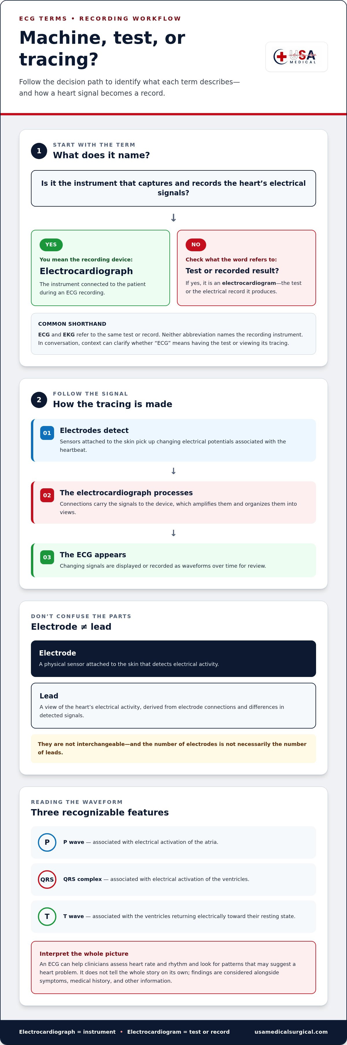
The key difference is what each term names: an electrocardiograph is the instrument that records the heart’s electrical activity, while an electrocardiogram is the test or the record it produces. Put simply, the machine makes the recording; the recording is the result. This distinction makes the terms electrocardiogram and electrocardiograph easier to understand without getting lost in technical language.
Quick definition: An electrocardiograph captures electrical signals from the heart and produces an electrocardiogram, often called an ECG or EKG. The recording provides information about the heart’s electrical activity, but it doesn’t diagnose every heart condition on its own.
Is an electrocardiograph the same as an electrocardiogram?
No. During an ECG recording, the electrocardiograph is the device connected to the patient, and the electrocardiogram is the resulting tracing or record. The test describes the recording process; the tracing shows the electrical signals captured during it.
In everyday conversation, a clinician or patient may say “ECG” to mean either having the test or looking at its tracing. The context usually makes clear whether they mean the procedure, the device, or the recorded result. For a broader overview of how these terms fit into the process, see Electrocardiography.
What do ECG and EKG stand for?
ECG is an abbreviation for electrocardiogram, based on the English spelling. EKG refers to the same test and record, but its “K” comes from the German-derived spelling Elektrokardiogramm. The alternate abbreviation doesn’t mean a different kind of test or change what the tracing represents.
Neither abbreviation names the recording instrument. That device is the electrocardiograph. You may see ECG or EKG used for the test or its output in clinical notes, patient instructions, and everyday discussion. The surrounding sentence usually indicates which meaning is intended.
Keep the distinction practical: the electrocardiograph is the equipment, and the electrocardiogram is what it records. ECG and EKG are common abbreviations for the test or its resulting record.
How an Electrocardiograph Turns Heart Signals into an ECG Tracing
An ECG tracing begins with small electrical differences detected at the skin. During a recording, electrodes placed on the body pick up these signals and connect to the electrocardiograph. The device processes and amplifies the signals so they can be displayed or recorded as a tracing over time.
An ECG device turns electrical signals detected at the skin into a visible tracing by receiving, amplifying, and recording them.
What role do ECG electrodes and leads play?
Electrodes are sensors attached to the skin. They detect electrical activity associated with the heartbeat, and wires or other connections carry the signals to the electrocardiograph for recording.
A lead is a view of the heart’s electrical activity, derived from connections between electrodes and differences in the signals they detect. An electrode is a physical sensor; a lead is a way of representing electrical activity. The terms aren’t interchangeable, and the number of electrodes isn’t necessarily the number of leads.
How does the signal become a tracing?
The process can be understood in a few steps:
- Electrodes detect signals: Sensors on the skin pick up changing electrical potentials.
- The device receives and processes them: The electrocardiograph amplifies the small signals and organizes them into views.
- The ECG is displayed or recorded: The device presents the changing signals as waveforms over time for review.
For a patient-focused overview of what to expect during the test, MedlinePlus on Electrocardiogram explains the recording process and how clinicians use the results.
What do the P wave, QRS complex, and T wave represent?
On a standard ECG tracing, the P wave, QRS complex, and T wave are recognizable waveform features. In general, the P wave is associated with electrical activation of the atria, the QRS complex with activation of the ventricles, and the T wave with the ventricles returning electrically toward their resting state.
These features help clinicians assess the heart’s electrical activity, but a waveform shouldn’t be interpreted in isolation. Its meaning depends on the full tracing and clinical context. Understanding how the electrocardiogram and electrocardiograph work together also clarifies the role of each part in the recording workflow. For settings that use this equipment, explore ECG machines and electrodes.
ECG, EKG, Cardiogram, Leads, and Electrodes: Which Terms Differ?
These terms describe different parts of an ECG recording, so they aren’t interchangeable. The electrocardiograph is the device, while the electrocardiogram is the test or its recorded result. ECG and EKG are common abbreviations for the test or tracing. The other terms refer to a heart-related record, the sensors that contact the skin, or the views used to represent electrical activity.
| Term | What it means |
|---|---|
| Electrocardiogram | The ECG test or the electrical record it produces. |
| Electrocardiograph | The instrument that captures and records electrical signals. |
| ECG or EKG | Abbreviations commonly used for the test or its tracing. |
| Cardiogram | A general or sometimes imprecise term for a record of heart activity. |
| Electrode | A sensor placed on the skin to detect electrical signals. |
| Lead | A view of cardiac electrical activity derived from electrode connections and signal differences. |
The device and recording are connected, but they are not the same thing. A clinician might say they’re reviewing an ECG, meaning the tracing, while the electrocardiograph is the equipment used to obtain it. Knowing the difference between electrocardiogram and electrocardiograph makes notes and equipment descriptions easier to interpret.
Is an ECG the same as a cardiogram?
Usage of “cardiogram” varies, so context matters. It may be used informally to mean a heart-related recording, but it isn’t always a precise name for an ECG. An ECG records electrical activity. An echocardiogram, by contrast, uses ultrasound to create images of the heart. Each provides different information, and neither alone captures every aspect of heart function.
Are ECG leads the same as electrodes?
No. Electrodes are the skin-contact sensors; leads are electrical views formed from signals detected through electrode connections. A useful distinction is sensor versus viewpoint, not one electrode per lead. The terms describe related parts of the recording setup, but the number of electrodes and leads shouldn’t be treated as interchangeable.
For details on device features and selection considerations, see the ECG machine buying guide. For practical positioning information, refer to the 12-lead ECG placement guide.
What an ECG Tracing Can Show, and What It Cannot Show Alone
An ECG records the heart’s electrical activity during the test. Clinicians can review the tracing for information such as heart rate, rhythm, and electrical patterns. It offers a time-limited view of electrical signals, not a complete picture of heart function.
An ECG doesn’t directly create images of the heart or measure blood flow. Other assessments may be needed to examine structures, pumping function, or circulation. The electrocardiogram and electrocardiograph are important parts of the recording process, but the tracing is evidence for clinical interpretation, not a diagnosis by itself.
Clinicians consider ECG findings alongside relevant information, including:
- The person’s symptoms and medical history
- Findings from a physical examination
- Other test results or clinical details, when appropriate
Can an electrocardiogram diagnose a heart condition by itself?
An ECG can contribute useful evidence, but it isn’t a complete clinical assessment. A finding may help guide further evaluation, while a normal-looking tracing doesn’t rule out every heart problem. A qualified healthcare professional interprets the result in context. If you have symptoms or concerns about your heart, discuss them with a healthcare professional rather than relying on an ECG reading alone.
Why can ECG results vary between recordings?
Some differences come from how the recording is captured. Electrode contact, body movement, and other recording conditions can affect signal quality. Timing matters, too: an ECG captures electrical activity during a particular period, and patterns may change over time or with a person’s clinical circumstances. A difference between recordings doesn’t explain its own cause, so trained staff and clinicians interpret it in context.
Understanding the test’s limits helps keep its role clear: an ECG can show electrical patterns, but it cannot answer every question about the heart. If you’re equipping a clinical setting, browse ECG machines and electrodes for the recording components used in an ECG workflow.
How Electrocardiographs Fit into Clinical Recording and Equipment Selection
An ECG recording follows a practical sequence: prepare the patient, position the electrodes, capture the tracing, then route the record for clinical review. Each part has a distinct role. Electrodes contact the skin and detect electrical signals; the electrocardiograph receives and records those signals as an ECG tracing.
ECG machines and ECG electrodes are separate equipment categories used together in this process. The recording device handles signal acquisition and presents or stores the resulting record, depending on its functions. Clinical interpretation remains the responsibility of a qualified clinician, not the machine alone.
What does an ECG machine do during a recording?
During a recording, an ECG machine receives electrical signals from the connected electrodes and processes them into a tracing. The tracing may be displayed or recorded for review. Device functions vary, so the way a record is presented or stored depends on the equipment. The machine captures and organizes the signal; clinicians interpret what the tracing may mean in the relevant clinical context.
When should readers explore ECG machines or electrodes?
Equipment needs depend on the intended setting and recording requirements. A clinical workflow may involve selecting a recording device and compatible electrodes as separate components of the setup. Electrodes support signal capture, while the electrocardiograph records the resulting signals. This distinction helps identify which part of the workflow you’re considering without treating the equipment as interchangeable.
For more detailed purchasing considerations and device-selection factors, consult the ECG machine buying guide. It covers selection in greater depth while explaining the distinct roles of the machine and electrodes.
Once you understand how the components fit together, browse the ECG equipment catalog to explore ECG machines and electrodes. Thinking of the electrocardiogram and electrocardiograph as the record and the recording instrument makes the equipment workflow clearer.
Put ECG Terms into Practice
The distinction is straightforward: the electrocardiograph is the instrument, and the electrocardiogram is the test or recorded tracing. ECG and EKG are common abbreviations, while electrodes and leads describe separate parts of the recording process. Keeping these terms clear makes it easier to understand how a tracing is captured and discussed.
An ECG records electrical activity during the test. Clinicians may assess rate, rhythm, and electrical patterns, but the tracing doesn’t directly image the heart or measure blood flow. Its findings need to be interpreted alongside symptoms, medical history, and other relevant information.
Understanding how the electrocardiogram and electrocardiograph relate can also make equipment needs easier to follow. ECG machines record the signals, while ECG electrodes support signal capture. USA Medical and Surgical Supplies’ online catalog includes both categories.
Browse ECG machines and electrodes to explore equipment used in the recording workflow. With the terminology clear, you can approach ECG information and equipment with greater confidence.
Frequently Asked Questions
What is the difference between an electrocardiogram and an electrocardiograph?
An electrocardiograph is the instrument that records the heart’s electrical signals; an electrocardiogram is the test or the record it produces. For example, a clinician uses an electrocardiograph to capture a patient’s signals, then reviews the resulting ECG tracing. People often use “ECG” as shorthand for either the test or its record. The terms electrocardiogram and electrocardiograph refer to connected parts of the process, not the same thing.
Is an electrocardiograph the same thing as an ECG machine?
Yes. “Electrocardiograph” is the technical term for the device commonly called an ECG machine. It receives electrical signals from electrodes placed on the body and presents or records them as a tracing. The machine captures the information, but it doesn’t replace clinical interpretation. The recorded result is the electrocardiogram, also commonly called an ECG or EKG.
What does an electrocardiogram measure?
An electrocardiogram records the heart’s electrical activity during the recording period. Clinicians can review the tracing to assess features such as heart rate, rhythm, and electrical patterns. An ECG doesn’t directly image the heart or measure blood flow. It provides one type of information, which a clinician interprets alongside symptoms, medical history, examination findings, and other relevant results.
Are ECG and EKG the same test?
Yes. ECG and EKG are two abbreviations for the same type of test and its recorded tracing. ECG comes from the English spelling of electrocardiogram, while EKG reflects the German-derived spelling Elektrokardiogramm. The different letters don’t indicate different tests. In clinical notes or conversation, context usually shows whether ECG or EKG refers to having the test or reviewing its result.
Can an ECG show whether a heart is healthy?
An ECG can provide useful information about the heart’s electrical activity, but it can’t establish overall heart health by itself. A normal tracing doesn’t rule out every heart condition, and an unusual pattern needs clinical interpretation. ECG findings are considered with symptoms, medical history, examination, and other appropriate information. If you have concerns or symptoms, discuss them with a qualified healthcare professional rather than relying on a tracing alone.
What is the difference between ECG leads and electrodes?
Electrodes are sensors that make contact with the skin and detect electrical signals. Leads are views of the heart’s electrical activity derived from connections between electrodes and differences in the detected signals. Electrodes are physical parts of the setup, while leads describe how the electrical information is represented. The number of electrodes and the number of leads aren’t interchangeable measures.
How does an electrocardiograph create a tracing?
Electrodes placed on the skin detect changing electrical signals and pass them to the electrocardiograph. The device receives and amplifies the signals, then displays or records them as waveforms over time. Those waveforms form the ECG tracing for review. Recording conditions and movement can affect signal quality, and a clinician interprets the completed tracing in context rather than treating the machine’s output as a diagnosis.