
Fetal Doppler Technology: Clinical Guide to Selection, Frequencies, and Diagnostic Use
09/25/2026 05:08:41

Selecting an advanced fetal doppler won't guarantee diagnostic accuracy if the probe frequency doesn't match your patient's gestational stage. Every practitioner knows the frustration of isolating faint acoustic signals, only to realize the unit picked up maternal blood flow instead of a true fetal heart rate between 110 and 160 beats per minute. Probe confusion between 2 MHz and 3 MHz configurations adds friction to routine exams, while uncertainty regarding device durability and regulatory compliance can complicate procurement decisions.
Reliable prenatal monitoring demands clear acoustic separation and dependable hardware built for daily clinical use. In this guide, you'll learn how continuous wave ultrasound technology functions in prenatal diagnostics, evaluate essential technical specifications, and identify the ideal device for your practice. We'll walk through probe selection across different trimesters, review key safety standards, and explain how to source durable diagnostic equipment from an authorized medical distributor.
Key Takeaways
- Master continuous wave ultrasound mechanics to isolate cardiac acoustic reflections and avoid maternal vascular interference.
- Match transducer frequencies to gestational stages, balancing early acoustic sensitivity with deeper anatomical penetration.
- Identify the core regulatory and build benchmarks that separate an authorized clinical fetal doppler from unregulated consumer monitors.
- Apply systematic abdominal mapping and acoustic coupling techniques to eliminate signal noise during routine auscultation.
- Standardize procurement by evaluating probe interchangeability, enclosure durability, and authorized distributor fulfillment.
Understanding Fetal Doppler Technology: How Continuous Wave Ultrasound Detects Heartbeats
A handheld fetal doppler serves as a primary non-invasive diagnostic instrument in modern obstetrics. Clinical staff rely on these portable devices during routine prenatal visits to assess fetal viability rapidly between full ultrasound scans. Unlike standard stethoscopes, which rely on mechanical sound conduction, a clinical Doppler fetal monitor utilizes high-frequency sound waves to capture the physical motion of cardiac structures deep within the pelvic cavity.
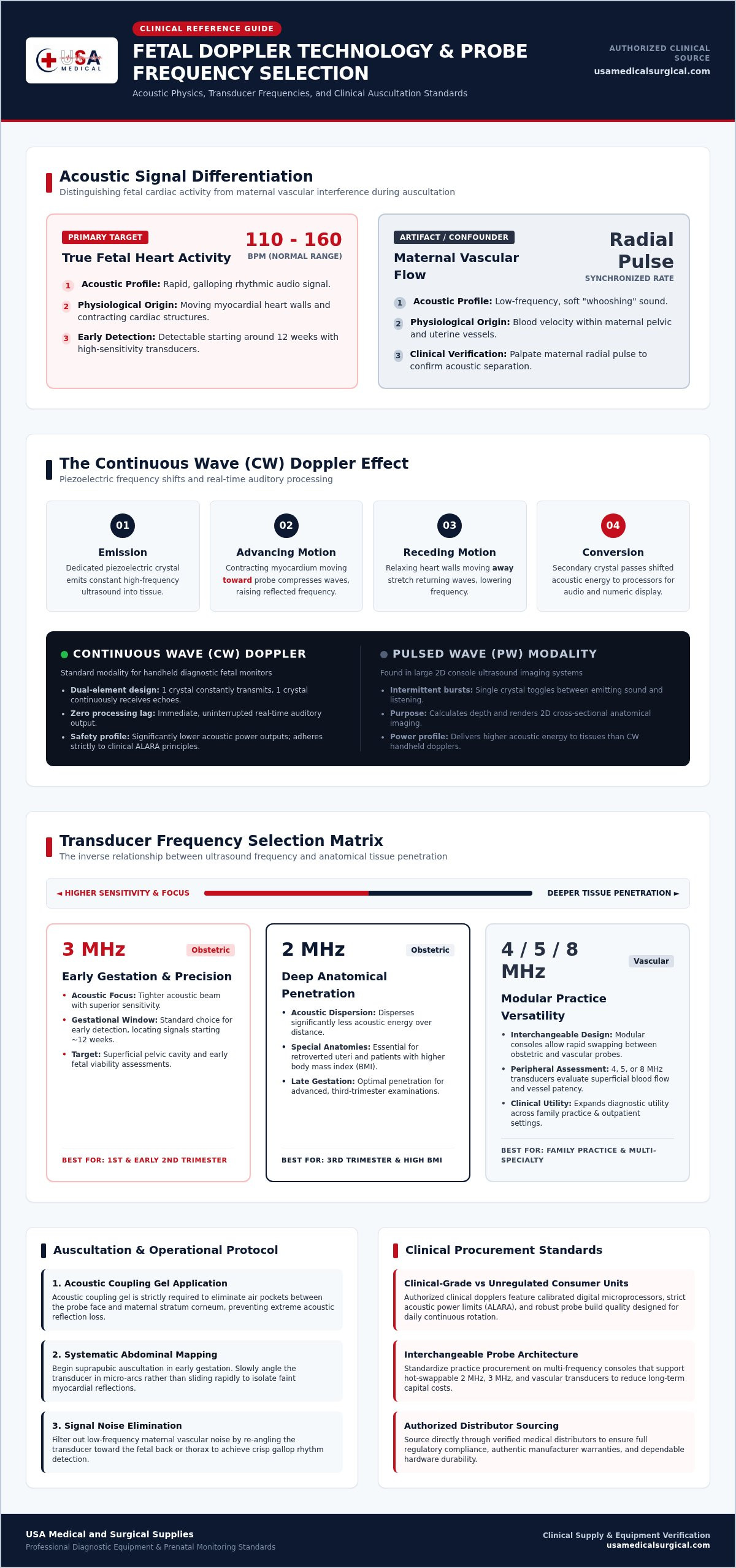
Accurate auscultation hinges on distinguishing true fetal cardiac activity from surrounding maternal vascular flow. Maternal pelvic vessels generate low-frequency, whooshing sounds synchronized with the patient's radial pulse. Fetal heart activity produces a distinct, rapid galloping rhythm, typically operating at 110 to 160 beats per minute. Diagnostic handheld units filter and amplify these frequency shifts, helping clinicians isolate true fetal signals without acoustic ambiguity.
The Physics of the Acoustic Doppler Effect
The device operates on acoustic wave reflection. Piezoelectric crystals housed within the probe face emit continuous sound waves into maternal abdominal tissue. When these waves encounter stationary structures, they bounce back at their initial frequency. When they strike moving red blood cells or contracting myocardium, the reflected frequency shifts:
- Advancing motion: Cardiac tissue moving toward the transducer compresses the returning waves, raising the frequency.
- Receding motion: Relaxing heart walls moving away stretch the waves, lowering the returning frequency.
- Signal conversion: Secondary receiver crystals capture this shifted acoustic energy and pass it to internal processors, which convert the shift into audible sound and real-time numerical data.
Continuous Wave Ultrasound vs Pulsed Diagnostic Modalities
Handheld fetal monitors employ continuous wave (CW) ultrasound rather than the pulsed wave (PW) systems found in large imaging consoles. In a continuous wave setup, one dedicated crystal transmits sound constantly while an adjacent crystal continuously receives echoes. This dual-element design provides a continuous, real-time auditory feed of cardiac wall motion without processing lag.
Pulsed systems emit intermittent sound bursts to measure depth and render two-dimensional anatomical cross-sections. While invaluable for structural evaluations, pulsed ultrasound delivers higher acoustic energy to tissues. Continuous wave units maintain significantly lower acoustic power outputs. This conservative power profile preserves patient safety, aligns with clinical ALARA principles, and delivers immediate acoustic confirmation during rapid outpatient evaluations.
Core Technical Specifications: Selecting Frequency, Probe Design, and Display Output
Matching hardware specifications to daily patient volume ensures fast, definitive examinations. While basic monitors offer static configurations, a clinical-grade fetal doppler provides modular options to handle anatomical variations across all three trimesters. Evaluating transducer physics, display technology, and cable build quality helps practices select instruments that hold up under continuous clinical rotation.
Ultrasound Probe Frequencies: 2 MHz vs 3 MHz
Acoustic physics dictates an inverse relationship between ultrasound frequency and tissue penetration. Selecting the correct probe frequency depends on gestational age and maternal body habitus:
- 3 MHz probes: Higher frequencies offer a tighter acoustic focus and superior sensitivity. This makes a 3 MHz transducer the standard choice for detecting early cardiac activity, often locating signals starting around twelve weeks.
- 2 MHz probes: Lower frequencies disperse less acoustic energy over distance. A 2 MHz transducer delivers deeper penetration, making it ideal for third-trimester examinations, retroverted uteri, or patients with higher body mass index.
- Interchangeable systems: Multi-frequency units let clinicians swap between obstetric probes and vascular probes (such as 4, 5, or 8 MHz transducers) on a single console, expanding utility across family practice settings.
Audio Signal Clarity and Digital Display Processing
Modern diagnostic units integrate onboard microprocessors that process raw frequency data into clean visual and auditory metrics. High-resolution backlit LCD screens display real-time fetal heart rate calculations alongside signal strength indicators, eliminating manual counting errors during busy rounds. Built-in digital auto-correlation algorithms continuously smooth out rate fluctuations for dependable record-keeping.
Acoustic filtering plays a critical operational role. Advanced processing suppresses background noise caused by probe movement, maternal intestinal motility, and placental blood flow. Clinicians can output sound through integrated high-output speakers for family presence or route audio through a dedicated 3.5 mm headphone jack during loud clinical environments. When configuring outpatient exam rooms, reviewing professional diagnostic Dopplers ensures your team secures units equipped with reliable digital processing.
Transducer Ergonomics and Waterproof Capabilities
Daily rounds involve repetitive probe manipulation, making transducer weight and balance essential considerations. Ergonomic contoured grips reduce practitioner hand strain during prolonged localization attempts. Heavy-duty coiled cables provide practical reach around examination tables without dragging across contaminated floor surfaces.
Sealed waterproof transducers, typically rated to IPX7 standards, offer two distinct practical benefits. They enable continuous acoustic auscultation during labor in water birth suites, and they withstand complete immersion in chemical disinfectants between patients. This seamless fluid resistance simplifies infection control routines and prevents internal crystal degradation over years of heavy practice use.
Clinical Diagnostic Dopplers vs Consumer Devices: Regulatory and Build Standards
Procurement teams and clinicians must draw a sharp line between professional diagnostic instruments and direct-to-consumer gadgets. Over-the-counter devices often present themselves as clinical solutions, but they lack the manufacturing consistency, safety testing, and signal accuracy required for medical decision-making. Investing in a professional fetal doppler ensures diagnostic reliability, legal compliance, and long-term equipment integrity.
FDA Oversight and Medical Device Classifications
The U.S. Food and Drug Administration classifies diagnostic fetal monitors as Class II medical devices under 21 CFR 884.2660. Bringing a clinical unit to market requires a comprehensive 510(k) premarket notification confirming substantial equivalence to established predicate hardware. In contrast, unregulated novelty devices frequently bypass medical clearance pathways entirely.
FDA standards establish strict limits on spatial-peak temporal-average acoustic output, requiring devices to adhere to international electrical safety (IEC 60601-1) and electromagnetic compatibility benchmarks (IEC 60601-1-2). International regulators take an even stricter stance. For instance, the Australian Therapeutic Goods Administration removed all home-use fetal monitors from the Australian Register of Therapeutic Goods in January 2026 due to safety risks from untrained interpretation. Professional devices provide calibrated acoustic energy that guarantees clinical safety throughout routine assessments.
Signal Artifacts: Distinguishing Fetal Pulse from Maternal Arteries
Untrained users and low-grade consumer units frequently struggle with signal ambiguity. Professional hardware incorporates sensitive digital processing to minimize false readings caused by adjacent anatomical structures:
- Fetal cardiac activity: Produces rapid, rhythmic sounds that mirror a galloping horse, typically holding between 110 and 160 beats per minute.
- Maternal vascular flow: Generates slower, sweeping whooshes from the uterine artery that sync with the patient's pulse.
- Placental souffle: Creates continuous, windswept acoustic sounds caused by blood rushing through expanded vascular channels.
Clinical-grade units feature advanced autocorrelation processing that tracks the true fetal rate while filtering out lower-frequency maternal pulses. This technical separation prevents false reassurance during critical assessments.
Infection Control and Enclosure Durability
Clinical workflows demand rugged physical hardware. Consumer devices rely on lightweight casing that fractures easily and degrades under chemical cleaning. Professional diagnostic units utilize high-impact ABS polymer housings engineered to survive accidental drops onto tiled clinic floors.
Hospital infection control requires relentless sanitization between every patient encounter. Clinical monitors feature sealed membrane buttons, liquid-resistant probe housings, and recessed display bezels. These barrier designs prevent conductive ultrasound gel, alcohol wipes, and quaternary ammonium disinfectants from seeping into internal circuit boards, preventing premature electronic failure in high-volume practices.
Operational Protocol: Transducer Placement, Coupling Gel, and Maintenance
Executing a consistent auscultation protocol minimizes examination time and prevents diagnostic frustration. Even a high-performance fetal doppler requires methodical handling to bypass acoustic interference from bowel gas and adipose layers. Establishing standard clinical workflows protects sensitive probe elements while maintaining clean signal acquisition across every shift.
Auscultation Technique Across Gestational Stages
Anatomical landmarks change dramatically as pregnancy advances, requiring targeted transducer positioning:
- Early second trimester (12 to 18 weeks): Place the probe face midline, immediately superior to the pubic symphysis. Angle the beam downward into the pelvic brim. Tilt the transducer face slowly in small radial arcs instead of sliding it across dry skin.
- Mid-gestation (18 to 28 weeks): Move the probe upward toward the lower umbilical region. Fetal mobility increases significantly here, requiring systematic four-quadrant searching if cardiac signals shift during localization.
- Third trimester (28 weeks onward): Palpate Leopold maneuvers first to confirm presentation and identify the fetal back. Positioning the transducer directly over the smooth convex plane of the fetal spine provides the shortest acoustic distance to the myocardium.
Acoustic Coupling Gel Application Guidelines
Ultrasound waves travel poorly through air. Placing a transducer against bare skin creates acoustic mismatch, reflecting over 99 percent of sound waves before they enter the patient. Applying a generous bead of aqueous acoustic transmission gel directly to the probe face bridges this boundary layer.
Never substitute ultrasound gel with surgical lubricants, hand lotions, or mineral oils. Petroleum-based products and cosmetic additives soften acoustic faceplates and degrade probe perimeter seals over time. Maintain moderate, even pressure against the abdominal wall. Lifting the probe breaks the acoustic bridge and floods the speakers with harsh, distorted static.
Device Sanitation and Transducer Care
Piezoelectric elements mounted within the probe housing fracture easily under mechanical stress. Always dock the transducer securely in its molded bracket between evaluations. To preserve equipment readiness, restock clinical examination suites with professional grade Dopplers and dedicated diagnostic supplies through an authorized national distributor.
Follow a disciplined cleanup routine after every patient encounter:
- Immediate gel removal: Wipe residual gel from the probe face immediately using soft, non-abrasive hospital cleaning wipes. Dried gel hardens into an acoustic barrier and harbor contaminants.
- Disinfection limits: Clean only the lower waterproof section of the probe assembly. Never submerge cable interconnect pins or console housings in liquid baths unless explicitly rated for immersion.
- Visual checks: Inspect the transducer lens weekly for hairline fractures. Cracked casings invite conductive fluid ingress that short-circuits internal components.
Procurement Standards: Sourcing Diagnostic Dopplers for Practices and Facilities
Outfitting an obstetric suite or outpatient women's health clinic requires a calculated purchasing strategy. Clinical buyers must look beyond upfront equipment costs to evaluate lifecycle reliability, parts availability, and daily maintenance demands. Sourcing a compliant clinical fetal doppler through proper commercial medical channels protects practice budgets while ensuring uninterrupted patient care across high-volume exam schedules.
Selecting Equipment Built for Clinical Longevity
Healthcare facilities require hardware built to endure continuous daily rotation. When evaluating potential devices for clinical suites, procurement teams should prioritize three key operational criteria:
- Modular components: Transducer cables experience significant physical strain over time. Select units featuring detachable, interchangeable probes so your facility can replace a worn cable or probe without discarding the primary base station.
- Power management: Battery depletion disrupts appointment pacing. Units equipped with drop-in desktop charging docks keep instruments fully charged between exams, eliminating the ongoing cost and waste of disposable alkaline cells.
- Manufacturer validation: Verify that hardware adheres to established diagnostic calibration standards, backed by clear manufacturer warranties and documented compliance certificates.
Procuring Reliable Diagnostic Instruments from USA Medical and Surgical Supplies
Sourcing diagnostic ultrasound monitors through open third-party marketplaces exposes facilities to counterfeit goods, gray-market imports, and unverified warranties. Partnering with a dedicated medical supply distributor guarantees direct access to authentic clinical equipment backed by manufacturer support.
Consolidating equipment and consumable orders simplifies clinical supply chains. In addition to primary diagnostic instruments, outpatient facilities require steady access to medical-grade transmission gel, specialized probe sanitizers, and compatible infection control supplies. Securing these supplies from a single vendor streamlines inventory management and reduces shipping delays. Contact USA Medical and Surgical Supplies for reliable nationwide diagnostic equipment, dependable supply fulfillment, and verified diagnostic instruments tailored to clinical practice needs.
Standardizing Prenatal Diagnostic Accuracy Across Your Facility
Selecting the right diagnostic tools directly impacts clinical workflow and exam efficiency. Sourcing a certified fetal doppler matched to patient gestational stages eliminates acoustic confusion, allowing your staff to differentiate maternal vascular flow from true fetal heart rates with confidence. Class II medical-grade hardware ensures your facility adheres to strict acoustic output limits, withstands routine chemical disinfection, and maintains calibration across high-volume schedules.
Outfitting your examination suites doesn't have to complicate clinical procurement. As an authorized distributor of diagnostic clinical equipment, USA Medical and Surgical Supplies delivers nationwide fulfillment across the United States. With a comprehensive catalog supporting practices, facilities, and caregivers, you can reliably acquire durable monitors, replacement transducers, and acoustic coupling supplies. Explore Clinical Fetal Dopplers and Diagnostic Instruments to equip your teams with dependable diagnostic technology for every prenatal exam.
Frequently Asked Questions
What is the primary difference between a 2 MHz and a 3 MHz fetal Doppler probe?
A 3 MHz probe provides higher acoustic frequency for early detection, while a 2 MHz probe provides deeper tissue penetration for later pregnancy or higher patient BMI. Higher frequencies scatter faster in tissue, making 3 MHz ideal from roughly 12 weeks. In contrast, 2 MHz waves travel deeper with less dispersion, making them reliable during the third trimester or when monitoring through deeper abdominal layers. Many facilities maintain both interchangeable options.
At how many weeks gestation can a fetal Doppler detect a heartbeat?
A clinical fetal doppler reliably detects a fetal heartbeat between 10 and 12 weeks of gestation. Auscultation earlier than 10 weeks is rarely achievable because the uterus remains deep within the pelvic cavity behind the pubic bone. Patient anatomy, placental position, and probe frequency also influence timing. If signals aren't detected by week 12, clinicians typically verify fetal viability and position using two-dimensional diagnostic ultrasound imaging consoles.
Can ultrasound transmission gel be substituted with regular lotion or oil?
No, you should never substitute ultrasound transmission gel with lotions, oils, or hand sanitizers. Dedicated aqueous gel eliminates air pockets between the probe face and skin, allowing seamless acoustic wave conduction. Lotions and petroleum-based products don't provide adequate acoustic impedance matching. Over time, cosmetic oils and chemical additives also degrade the protective acoustic membrane and perimeter seals on sensitive transducer heads, causing permanent hardware damage.
How do clinicians tell the difference between the mother's pulse and the fetal heart rate?
Clinicians differentiate the signals by rhythm cadence and simultaneous pulse verification. A fetal heart rate beats rapidly at 110 to 160 beats per minute, creating a sharp galloping cadence. Maternal pelvic blood flow produces a lower-frequency, swooshing sound matching the maternal pulse. Palpating the patient's radial pulse while listening instantly identifies maternal vascular signals, ensuring diagnostic clarity during routine examinations.
Are handheld fetal Dopplers safe for routine prenatal examinations?
Yes, clinical fetal doppler devices are safe for routine prenatal examinations when used by trained healthcare personnel. These instruments emit continuous wave ultrasound at very low acoustic output levels, complying with FDA Class II medical standards and international safety guidelines. Clinicians adhere to the ALARA (As Low As Reasonably Achievable) principle by minimizing examination duration once they confirm and document stable cardiac rhythm.
What causes static and audio interference during Doppler auscultation?
Static and audio noise stem primarily from poor acoustic coupling, rapid probe movement, or maternal bowel activity. Inadequate ultrasound gel creates microscopic air pockets that reflect sound waves abruptly back into the transducer. Moving the probe face too quickly across dry skin introduces friction noise. Additionally, gas movement in the intestines generates low-frequency rumble, which advanced digital audio filters help suppress during clinical exams.
What features differentiate professional clinical Dopplers from consumer units?
Professional monitors feature regulated acoustic outputs, durable high-impact ABS enclosures, calibrated digital rate calculation, and interchangeable waterproof transducers. They comply with strict FDA 510(k) and IEC 60601-1 standards to ensure electrical safety and calibration stability across years of daily use. Consumer novelties lack hospital-grade disinfection compatibility, sensitive noise filtration, and verified manufacturing tolerances, creating risks of false reassurance and inaccurate diagnostic readings.