
Ear Examination Devices for Practitioners: How to Choose a Scope
10/21/2026 09:01:25
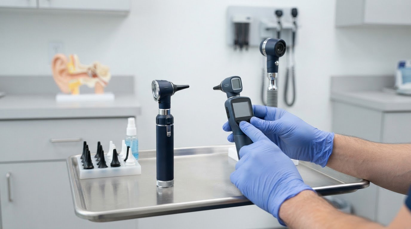
The right ear examination device for practitioners depends on the task. An otoscope lets practitioners examine the ear canal and eardrum. FDA registration may also matter, but it does not by itself show whether a scope’s viewing system, illumination, or portability suits your clinical setting.
Choosing a device means balancing familiar features with the demands of routine examinations. This guide explains the main otoscope types and what to compare, including magnification, viewing format, and use in a fixed office or across different settings.
You’ll also learn how otoscopes support ear assessment and where a digital model may fit in a practice. Use these practical considerations to narrow your options, including how to approach the FDA-registration question.
Key Takeaways
- Match the scope to the setting: office examinations and mobile use can call for different formats, including desktop, wall-mounted, or portable options.
- Compare how the instrument illuminates and presents the ear canal and eardrum, not just its overall design.
- Consider whether pneumatic otoscopy is relevant to the assessment, since it evaluates eardrum movement in response to pressure.
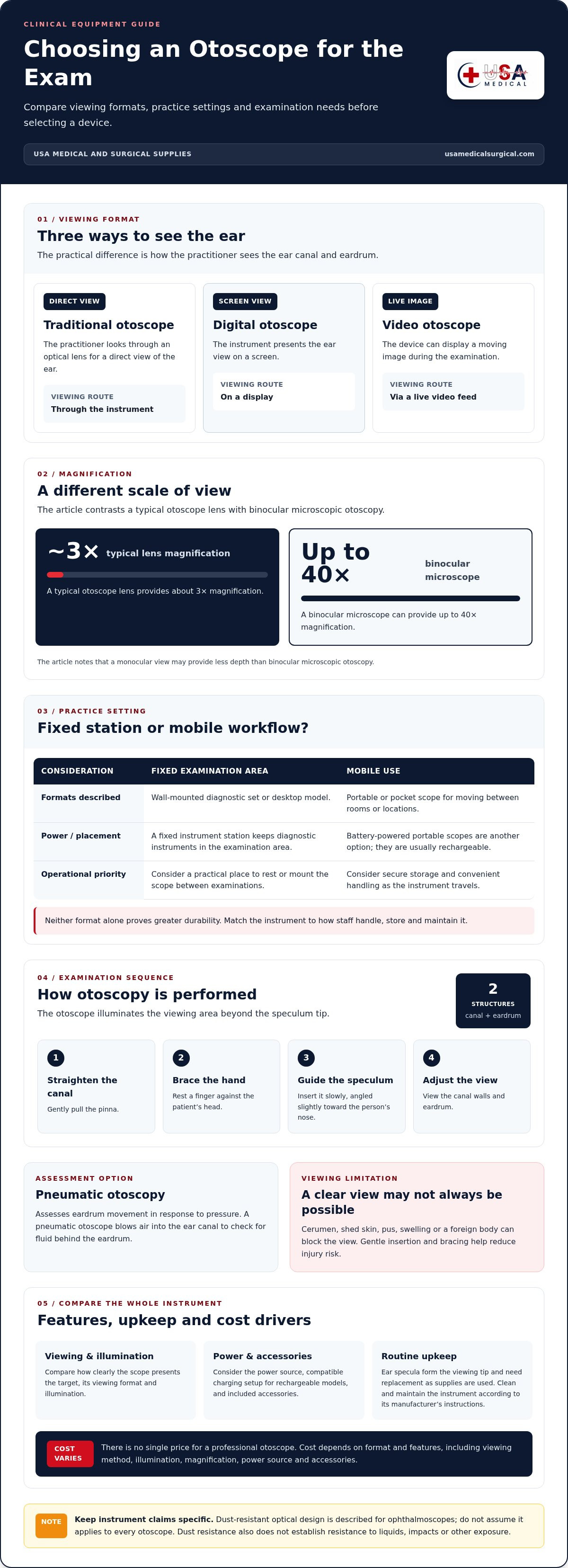
- Understand the viewing trade-off: a typical otoscope lens provides about 3× magnification, while a binocular microscope can provide up to 40×.
- Keep device features distinct: dust-resistant optical design is described for ophthalmoscopes, so assess durability based on the specific instrument.
Where Can Digital Otoscopes Be Used?
Practitioners use otoscopes in medical offices to examine the ear canal and eardrum. They can also support routine physical examinations, where clinicians assess the ears alongside other parts of the body. A digital model can serve in an office examination room or another clinical setting where viewing the ear is part of the work.
Traditional otoscopes provide a direct view through an optical lens. Digital otoscopes show the view on a screen, while video otoscopes can display a moving image during an examination. The practical difference is how the practitioner sees the ear: through the instrument, on a display, or via a live video feed.
Wall-mounted diagnostic sets keep instruments in a fixed examination area, while portable otoscopes suit clinicians who move between rooms or locations. Otoscope and ophthalmoscope sets are available as wall kits or desktop models. Battery-powered portable scopes, usually rechargeable, are another option for settings without a fixed instrument station.
During otoscopy, the practitioner examines the outer ear canal and eardrum. The otoscope’s light extends beyond the speculum tip to illuminate the viewing area. To perform the examination, the practitioner gently pulls the pinna to straighten the canal, braces a finger against the patient’s head, and slowly guides the speculum into the ear. The speculum angles slightly toward the person’s nose; the examiner adjusts the otoscope to view the canal walls and eardrum.
There is no single price for a professional otoscope. Cost depends on the format and features, such as optical viewing versus a digital display, illumination, magnification, power source, and included accessories. When comparing scopes, consider how clearly each presents the target, whether it suits the examination setting, and whether its power and handling work for regular use.
Common accessories include ear specula, which form the viewing tip; replacement specula are needed as supplies are used. Battery-powered scopes also rely on their power components, and rechargeable models need a compatible charging setup. Clean and maintain the instrument according to the manufacturer’s instructions, including care for the lens, handle, and reusable parts. Use the cleaning and disinfection methods specified for that device rather than assuming a method is suitable.
Otoscopy has limits when cerumen, shed skin, pus, swelling, or a foreign body blocks the view. A monocular view may also provide less depth than binocular microscopic otoscopy. Pneumatic otoscopy assesses eardrum movement in response to pressure. According to the National Institute on Deafness and Other Communication Disorders, a pneumatic otoscope blows air into the ear canal to check for fluid behind the eardrum. Gentle insertion and bracing help reduce injury risk during the examination.
Durability and dust-resistant design
Durability depends on how an otoscope is handled, stored, and cleaned in its intended setting. A pocket scope may travel between examination rooms, while a full-size instrument may stay at a fixed workstation. These routines call for different priorities: a mobile device needs secure storage and convenient handling; a stationary scope needs a practical place to rest or mount between examinations. Neither format alone proves greater durability.
Dust protection is a specific design feature, not a general measure of ruggedness. Dust-resistant designs are described for ophthalmoscopes to protect optical components. That claim applies to the named instrument category and should not be assumed to describe every otoscope. Dust resistance also does not establish resistance to liquids, impacts, or other exposure. Assess each feature for the specific instrument and its intended use.
Compare construction details that affect routine handling instead of relying on labels such as “professional” or “heavy-duty.” Check how the head connects to the handle, whether controls are easy to operate without awkward gripping, and how the scope can be stored when not in use. For a pocket model, consider whether it can be carried securely without its lens or controls pressing against other equipment. For a full-size model, consider how its stand or wall position protects it from accidental knocks.
- Optical components: Check whether the lens and viewing area have protection suited to the storage environment.
- Handle and controls: Consider grip, switch placement, and how the instrument is held during repeated examinations.
- Storage: Match the case, holder, or fixed location to whether the scope travels or remains in one room.
- Cleaning routine: Follow the instrument manufacturer’s instructions, especially for parts that must be cleaned after use.
Maintenance can preserve an instrument’s usable condition, but it cannot change its design. A carrying case may reduce contact with other items, but it does not demonstrate impact resistance. Likewise, a dust-resistant component does not mean the whole instrument can be exposed to dust without limits. Store the scope as intended, handle optical parts carefully, and follow the manufacturer’s cleaning guidance for the specific model.
For a primary care practice, the useful comparison is operational: a pocket scope suits a workflow that requires portability, while a full-size scope may better fit a dedicated examination station. Choose the form that supports the way staff handle, store, and maintain the instrument. Explore otoscope options alongside the durability needs of your practice.
Clean the otoscope according to the manufacturer’s instructions, especially any parts that need cleaning after use. Handle optical components carefully, store the instrument in its intended place, and follow model-specific guidance for disinfection and maintenance.
Ear canal and eardrum assessment
An otoscopic examination lets a practitioner inspect the outer ear canal and eardrum. It can help assess earache, a feeling of fullness, or hearing loss. Findings may involve the canal, eardrum, or middle ear. The otoscope’s light extends beyond the speculum tip to illuminate the area viewed through the instrument.
Technique affects the view and helps reduce the risk of injury. The practitioner gently pulls the pinna to straighten the canal, then braces a finger against the patient’s head. The speculum enters slowly and angles slightly toward the person’s nose, following the canal’s path. Small adjustments to the otoscope’s angle help reveal the canal walls and eardrum without forcing the tip forward.
Assessment depends on what the view reveals. Cerumen, shed skin, pus, swelling of the canal skin, and foreign bodies can obscure the eardrum. When these block the sightline, a common otoscope may not provide a clear view of the membrane and nearby structures. In practice, addressing heavy cerumen buildup often requires specialized care, where independent clinics like Ear and Hearing utilize microsuction to safely clear the canal. A larger viewing area can help practitioners inspect the ear, but a clear view still depends on visibility within the canal.
A standard monocular otoscope provides a single-eye view. Compared with binocular microscopic otoscopy, reliance on a monocular otoscope to diagnose ear disease results in a more than 50% chance of misdiagnosis. The comparison highlights a practical limitation: the view produced by a handheld scope may not provide the same depth as binocular microscopic examination. The instrument supports assessment, but its viewing format shapes what the examiner can observe.
Pneumatic otoscopy adds a functional assessment of eardrum mobility. The examiner uses positive and negative pressure to observe the membrane’s movement. According to the National Institute on Deafness and Other Communication Disorders, a pneumatic otoscope blows air into the ear canal to check for fluid behind the eardrum. A pneumatic otoscope is the standard tool used to diagnose otitis media.
These differences matter when selecting a scope for primary care. Consider whether routine visual inspection meets your examination needs, or whether assessing eardrum movement is also part of the work. For suspected acute otitis media, traumatic eardrum perforation, or cholesteatoma, otoscopic examination can contribute to diagnosis. The practitioner’s view, the structures visible, and the examination method all inform the assessment.
During an examination, the practitioner looks through the otoscope while slowly inserting the ear speculum into the canal. Otoscopy may help evaluate earache, a feeling of fullness, or hearing loss. Professional otoscopes are described as having a larger viewing area intended to help practitioners diagnose ear infections and diseases. Together, these points connect instrument use, symptoms it may help evaluate, and the diagnostic purpose of a larger viewing area.
Magnification for viewing ear structures
An otoscope’s magnifying lens helps practitioners inspect details inside the ear canal. A typical otoscope lens is around 8 diopters, providing approximately 3× magnification. That view supports routine examination through a handheld instrument. It can help reveal canal features and the tympanic membrane, but magnification alone doesn’t determine how clearly the practitioner sees them.
Enhanced magnification can help practitioners distinguish anatomical details and recognize foreign bodies in the ear canal and tympanum. For example, a small object may be easier to identify when enlarged through the optical view. Its usefulness still depends on illumination, lens clarity, positioning, and whether the canal is visible. Cerumen or swelling can obstruct the view regardless of magnification.
A binocular microscope used for otoscopy can provide up to 40× magnification. This is a different viewing approach from a monocular handheld otoscope. Greater magnification may support close inspection of structures, while a handheld scope offers a direct view through its lens. The figures describe magnification capacity, not a guarantee that every examination will reveal a particular finding.
For primary care, choose magnification to suit the examinations performed and the instrument’s handling. Consider whether the view makes it practical to assess the canal and eardrum during routine visits. Higher magnification can help reveal fine detail, but practitioners also need adequate illumination and a workable viewing area. A narrow, highly enlarged view may make orientation harder if the surrounding anatomy is difficult to locate.
- Magnification: Compare the stated lens power and magnification, such as the typical 8-diopter, approximately 3× otoscope lens.
- Viewing method: Distinguish a monocular handheld view from binocular microscopic otoscopy, which can reach up to 40×.
- Target detail: Consider whether the examination requires viewing general canal anatomy or identifying smaller features, such as a foreign body.
- Visibility: Account for illumination and obstructions that can limit the view, including cerumen or canal swelling.
Magnification is only one factor in selecting an ear examination scope. Match the optical view to the detail required, then consider how consistently staff can position and use the instrument. When comparing pocket and full-size otoscopes, assess each model’s viewing system alongside its form factor. The most useful choice supports clear inspection of the intended structures without treating magnification as a substitute for examination technique.
The viewing assembly matters too: an otoscope has a light, a magnifying lens, and a funnel-shaped viewing piece with a narrow, pointed end called a speculum.
What types of otoscopes are available for healthcare practitioners?
Compare otoscopes by viewing method, size, power source, and mounting format. These categories can overlap: a portable scope may use traditional optics, while a diagnostic set may combine an otoscope and ophthalmoscope. Start with the practice setting, then consider how staff will carry, position, and use the instrument. An ear examination device for practitioners should suit the actual workflow, not just a preferred form factor.
Pocket otoscopes are compact options for clinicians who move between rooms or work away from a fixed station. Portable otoscopes powered by batteries, usually rechargeable, are available. Their smaller format supports mobility, while the model’s controls, illumination, and lens shape the examination experience. Consider where the scope will be stored and how staff will recharge it during routine use.
Full-size otoscopes suit practices that prefer a larger handheld instrument at a regular examination station. Size alone does not establish optical performance or durability. Compare each model’s head, handle, viewing lens, and power arrangement. In a shared primary care room, consider where staff will place the instrument between examinations and how easily they can access it.
Traditional, digital, and video models differ in how they present the examination view. A traditional scope uses an optical viewing system. Digital and video designs use electronic viewing, with display features varying by model. Consider whether direct viewing or a screen-based image better fits the examination process. Do not assume a digital label guarantees a particular capability.
Pneumatic otoscopes support assessment of eardrum mobility using pressure. They are relevant when an examination requires more than visual inspection. Consider this type when evaluating eardrum movement is part of the practice’s clinical work, rather than assuming every scope includes pneumatic function.
Mounting and set formats offer another distinction. Otoscope and ophthalmoscope diagnostic sets are available as wall kits or desktop models. A wall kit keeps instruments at a designated station, while a desktop model sits on a work surface. A portable scope suits clinicians who carry equipment between locations.
- Choose a pocket format when portability matters to the workflow.
- Choose a full-size format when the instrument will mainly serve a fixed examination room.
- Compare viewing methods to match optical or screen-based viewing to the examination routine.
- Consider pneumatic function when assessment of eardrum mobility is relevant.
- Compare wall kits and desktop sets for a designated examination station.
For a practice selecting an ear examination device for practitioners, match the category to the examination method and workspace. Compare available otoscopes for clinical use by format and intended function.
USA Medical and Surgical Supplies: Blood Pressure Devices
USA Medical and Surgical Supplies offers Blood Pressure Devices for practices sourcing equipment beyond otoscopes. After choosing a scope format for your examination room or mobile workflow, browse equipment for other routine diagnostic needs.
Available categories include Blood Pressure Devices, Dopplers, ECG Machines, and ECG Electrodes. These products support different measurement and monitoring tasks, so choose according to your practice’s equipment needs. Browsing across categories can help when organizing purchases for several diagnostic needs, rather than selecting an otoscope alone.
Visit USA Medical and Surgical Supplies to browse available equipment.
Choose a Scope That Fits Your Next Step
Use your practice’s examination workflow as the final filter. Consider who will use the instrument, where it will be stored, and which viewing approach supports the assessments you perform. An ear examination device for practitioners should suit routine use as well as the specific needs of the clinical setting.
After settling on the scope requirements, plan related equipment needs separately. USA Medical and Surgical Supplies offers Blood Pressure Devices, Dopplers, and ECG Machines for practices sourcing diagnostic equipment across categories.
For help with your next equipment step, contact USA Medical and Surgical Supplies. Move forward with a scope choice grounded in how your practice works.
Frequently Asked Questions
Are These Devices FDA Registered?
FDA status depends on the specific otoscope, not just its category or intended clinical use. “FDA registered” alone does not establish that a particular model has the regulatory status your purchasing process requires. Identify the manufacturer and exact model, then review its labeling and applicable FDA device records. Do not infer registration, clearance, or approval from a product description alone.
What is an otoscope and how is it used in clinical practice?
An otoscope is a handheld diagnostic instrument for viewing the ear. In primary care, its findings can contribute to the clinical record alongside the patient’s symptoms and other examination results. For example, note which ear was examined, whether the eardrum was visible, and any relevant observations. Recording limits to the view helps distinguish an incomplete examination from a clearly observed finding.
Which otoscope brands and models are commonly used by practitioners?
Practitioners use different otoscope formats; no single model suits every setting. Common choices include pocket and full-size instruments, with optical, digital, or video viewing approaches. Compare how each model fits the practice’s workflow, including who will use it, where it will be stored, and how its view will be reviewed. Brand familiarity alone does not establish suitability.
What training and steps are recommended for performing an ear examination with an otoscope?
A practical training plan combines device familiarization, supervised technique, and consistent documentation. Practitioners can rehearse handling and positioning before examining patients, then review how to record the side examined, visible structures, and any limits to the view. Training should also cover when an examination needs further clinical assessment, rather than treating the otoscope image as a standalone diagnosis.
What U.S. regulations or standards apply to medical otoscopes?
Applicable U.S. requirements depend on the exact device and its intended use, so do not assume one regulation or standard covers every otoscope. For procurement, review the manufacturer’s intended-use statement, labeling, model-specific FDA status, and any standards the manufacturer identifies as applicable. A “professional,” “digital,” or “clinical” description alone does not establish regulatory compliance.
How much does a professional otoscope cost in the United States?
There is no single price for a professional otoscope; cost depends on its format and features.